

Vibrational spectroscopy
Infrared spectroscopy
In infrared (IR) spectroscopy the sample is irradiated with polychromatic light and a photon of light is absorbed when the frequency (energy) of the absorbed light matches the energy required for a particular bond to vibrate within the sample. For a vibration to be IR-active the molecular dipole moment must change during the vibration.
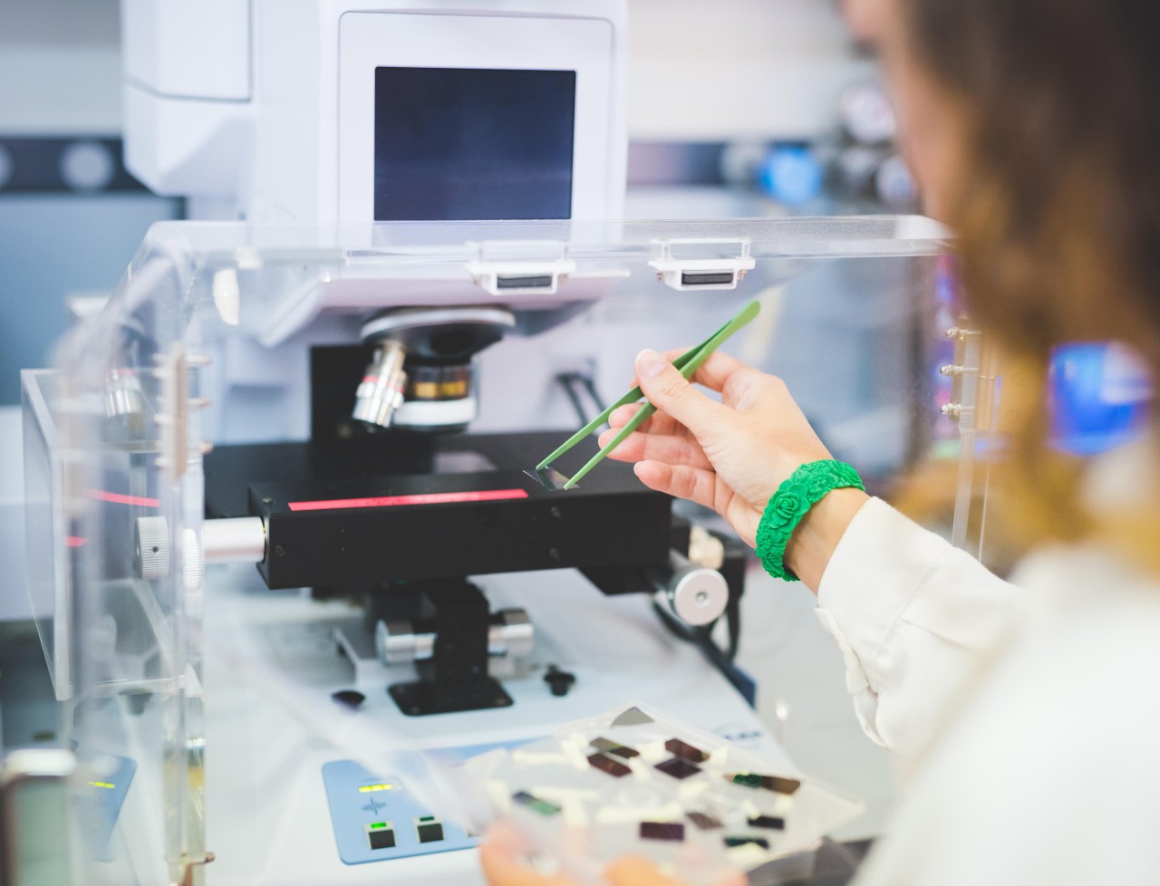
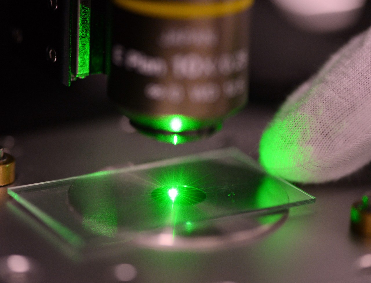
The Photothermal mIRage-LS system is a fully automated, sub-micron, multi-modal microscope platform that integrates Raman and Optical Photothermal Infrared (O-PTIR) spectroscopy with fluorescence microscopy. The instrument is capable of O-PTIR and Raman spectroscopy signal simultaneously from the same spatial location.
The system is equipped with dual Raman probe lasers at 532 nm and 785 nm, covering a spectral range of 4200 to 300 cm⁻¹. For infrared measurements, it utilises four Quantum Cascade Lasers (QCLs) spanning two spectral ranges: 2990–2700 cm⁻¹ and 1800–800 cm⁻¹. The spatial resolution of the system is approximately 500 nm and is independent of the IR wavenumber.
The mIRage-LS is suitable for a wide range of applications and material types, making it a versatile tool across various fields, from the study of microplastics, biological cells and tissues, materials in geological sciences to museum objects and archaeological artefacts.
The microtiter plate extension (HTS-XT module) enables high throughput screening of samples in transmission mode using standard microplate formats. Around 1 - 20 uL of the liquid sample is placed onto a single position of a 96-well silicon microtiter plate and dried prior to insertion into the spectrometer. A Twister Microplate Handler enables automatic loading and unloading of up to 20 microtiter plates for high-throughput analysis.
This instrument is also equipped with a Bruker BioATR II Unit that is specifically designed for the investigation of temperature-induced unfolding, refolding and denaturation processes of proteins in aqueous media using only 10-20 µL of sample. A programmable thermostat can produce temperatures from 5 to 95°C and can be set up for automatic ramping. The option for flow-through analysis is also available.
The Bruker Tensor 27 spectrometer is equipped to collect spectra over the MIR spectral range (7000 - 600 cm-1). It is coupled to the Hyperion 3000 microscope which has an MCT (Mercury Cadmium Telluride) detector allowing collection of spectra in single point and mapping modes.
Mapping involves the collection of spectra from specific, user-defined regions of interest using points, lines or grids. 2D/3D false colour maps are generated by plotting the intensity/area/peak-ratio of a characteristic peak(s) vs. the spatial x, y co-ordinates to obtain a visual distribution of one or more characteristic functional groups.
Specialised objectives are available for attenuated total reflectance (ATR) and grazing angle (GA) experiments and a live cell incubator accessory enables real-time analysis of changes in live cells under culture conditions.
In addition to standard transmission and reflection measurements, this system also has:
- Attenuated total reflectance (ATR) - used for analysis of materials that are strong absorbers and can provide information about the surface properties of a material
- Grazing angle (GA) - directs infrared light across the sample surface at an incident angle of ~ 84° creating an increased path length of the sample provide high sensitivity and making it ideal for microanalysis of thin coatings on metallic substrates, even down to monolayer thickness.
- Bioptech live cell incubator accessory - enables real-time analysis of changes in live cells under culture conditions.
- Linkam variable temperature stage – enables analysis of liquids or solids at temperatures ranging from –196° C to 600° C. There is also the option to have gas flow-through in the sample chamber.
This is a high-end research grade FTIR instrument equipped to collect spectra over the NIR (15500 – 4000 cm-1), MIR (12000 – 850 cm-1) and FIR (680 – 50 cm-1) spectral ranges using an evacuated optics bench. A vacuum pump allows for evacuation of the optic bench to significantly reduce the infrared active atmospheric water and carbon dioxide absorptions.
Other advantages that the vacuum system offers include the following:
- weak spectral features of some samples are not masked by moisture absorptions
- no problems are caused by the fluctuation of a dry air purge supply.
Sample accessories can be used with this spectrometer to enable analysis using the following method:
- diffuse reflectance – good for non-reflective materials (highly opaque or weakly absorbing) and irregular surfaces or coatings.
- specular reflectance – used to study film thickness, material reflectivity, and the uniformity and homogeneity of surface coatings.
- attenuated total reflectance (ATR) – used for materials that are strong absorbers and can provide information about the surface properties of a material.
- photoacoustic – suitable for solid, semi-solid and liquid samples as well as dark, highly absorbing samples.
This module interfaces with the Bruker Vertex 80v Spectrometer and provides information about the secondary structure of biomolecules (e.g. proteins, RNA/DNA) and how they change as a result of substrate binding, ligand binding, metal binding, oxidative stress, changes in pH or temperature; biomolecular docking and drug/target interactions.
- Samples are analysed in transmission mode as liquids or solids (pressed into pellets) over a spectral range of 1800-850 cm-1.
- Sum and difference signals are simultaneously collected with high sensitivity with the software able to perform calculation and normalisation of the VCD spectra.
- Short beam path provides optimal stability and signal-to-noise ratio.
This instrument provides morphological, mechanical property and IR spectral information on materials, surfaces and biomaterials (cells, tissues, extracellular vesicles, etc) at resolutions of less than 10 nm (for ideal samples). It consists of a NeaSNOM microscope with both a nano imaging module (NIM) and a nano-FTIR spectroscopy module (NSM). The scattering-type scanning near-filed optical microscope (s-SNOM) is based on atomic force microscope (AFM) technology where the spatial resolution is defined by the tip of the AFM probe.
The NIM uses a patented pseudo-heterodyne detection method coupled with a tuneable, single frequency QCL laser source to enable fast near-field image acquisition at a given wavelength within the range 1750 – 1500 cm-1 and 1200 – 915 cm-1. The second module, NSM, has two broadband lasers (4125 – 2400 cm-1 and 2250 – 667 cm-1) available for obtaining infrared spectra from individual points on a sample with a spectral resolution of up to 3.2 cm-1. This module is also capable of mapping measurements.
The instrument can handle samples up to 40 x 50 x 8 mm in size, while the x-y scanner can capture AFM and near-field images up to 100 µm x 100 µm with positioning resolution of 0.4 nm.
The system is also capable of photo-thermal expansion (PTE) spectroscopy and kelvin probe microscopy (KPFM).
This instrument is designed for the analysis of samples in the near-infrared (NIR) region (12500 – 3600 cm-1). It has high sensitivity and stability due in part to the permanently aligned RockSolidTM interferometer. A wide array of sampling methods makes it suitable for the analysis of liquids, solids, powders, pastes and tablets. The fibre optic probe attachment even allows samples to be measured directly without transferring to a sample holder. Transmission measurements of multiple liquid vials or solid tablets can also be carried out using the automated sample wheel.
A stand alone, fully automated, all-in-one microscope and FTIR spectrometer designed to be extremely easy to use while still providing high quality spectra. The instrument is fitted with a ×8 objective capable of measurements in reflectance, transmission and attenuated total reflectance (ATR) while a digital zoom allows visible light images to be collect at up to ×32 magnification. The numerical aperture of the objective is automatically changed when switching between IR and visible modes to provide high depth of field visible images while maintaining high sensitivity for IR analysis. The micro-ATR crystal has a 100 µm diameter and is fitted with an integrated pressure control to ensure consistent application of pressure by the crystal on the sample.
This is a standalone microscope offering fully automated measurements in all modes, the system is configured with three detectors, and it can undertake a range of experiments including point spectroscopy, mapping and imaging. The focal plane array (FPA) detector used to generate chemical images exceeding the speed and spatial resolution of line array detector. The LUMOS II is equipped with the following unique features:
- A standard TE-cooled MCT detector that does not require liquid nitrogen (6000-670 cm-1).
- A broadband liquid nitrogen cooled MCT detector (7800-450 cm-1)
- A 32x32 FPA imaging detector (4,000-750 cm-1)
- A long working distance of 30 mm and samples of up to 40 mm thickness can be measured.
- A built in 8x objective for transmission, reflection and retractable Ge (100 μm tip size) ATR objective.
- A dedicated Microplastics interface to automatically assign measurement spots and plastics identification.
- Contrast enhancement tools including polarisers and darkfield illumination.
The Spero-QT Chemical Imaging Microscope uses quantum cascade technology to substantially outperform FTIR microscopes in terms of spatial resolution, speed, and field-of-view capabilities, and does not require cryogenic cooling. The second-generation system has capability to produce twice the data in one-tenth of the time, while achieving unprecedented signal-to-noise ratios. The stage can image up to 3 microscope slides, and the large sample compartment makes the system compatible with microfluidic devices and accessories. ENVI software is used for advanced image analysis and data processing.
Partner facilities
Our partners at Australian Nuclear Science and Technology Organisation (ANSTO) operate two specialised IR beamlines at the Australian Synchrotron. We can provide advice on their suitability for your experiment and how to access them.
This is an FTIR spectrometer that uses the synchrotron as a source. Compared to conventional sources, synchrotron radiation spans a wider energy range (from microwave to hard X-rays) and is much brighter, collimated, and polarised (for circular dichroism and ellipsometry). This allows experiments on smaller samples and/or shorter timescales. For more information, see the beamline website.
This is an FTIR spectrometer that uses the synchrotron as a source, feeding into an IR microscope. The very higher intensity and collimation of synchrotron radiation permits measurement of microscopic samples at spatial resolutions of 3-8 μm, suitable for single-cell mapping. For more information, see the beamline website.
Raman spectroscopy
In Raman spectroscopy the sample is irradiated with monochromatic light and the photons are either inelastically or elastically scattered. The inelastically scattered light, known as Raman scatter, has lost (Stokes) or gained (anti–Stokes) energy during this interaction and the emitted photon contains information about the molecular structure of the sample. The elastically scattered light has the same energy as the incident laser light and is called Rayleigh scatter. Modern Raman instruments are designed to filter out the Rayleigh light as only one in every million photons will be Raman scattered. There is one other requirement for a vibration to be Raman active – when the molecule vibrates there must be a change in polarisability i.e., a change in the shape, size or orientation of the electron cloud that surrounds the molecules.
The microscope is a compact automated instrument used for a wide range of experiments from simple point spectroscopy to more sophisticated experiments. Multiple lasers offer excitation lines ranging from the visible to the near infrared (488, 514, 633, 785 and 830 nm) and provide the flexibility to offer the most suitable configuration for a researchers' experimental requirements.
The system is integrated with an atomic force microscope (AFM) to allow for correlated micro- and nano-scale property mapping using both AFM and tip-enhanced Raman spectroscopy (TERS).
Another feature of this system is the ability to map samples. Mapping is a specialised technique used to identify components and visualise their distribution within a sample. There are a number of different mapping modes available including: point-by-point, StreamLine™, StreamLine HR™, Surface and 3D Volume mapping.
Also available for use with this system are:
- Okolab live cell incubator stage - enables real-time analysis of changes in live cells under culture conditions.
- Linkam variable temperature stage – enables analysis of liquids or solids at temperatures ranging from –196° C to 600° C. There is also the option to have gas flow-through in the sample chamber.
The Renishaw InVia Qontor confocal spectrometer has an enclosed, upright microscope and features LiveTrackTM focus tracking technology that enables mapping of rough, smooth, flat and curved surfaces by automatically maintaining optimum focus during data collection. It is equipped with 532 and 785 nm excitation lines together with filters allowing for Raman and photoluminescence (PL) measurements to 50 cm-1 from the laser line. StreamHR™ Rapide enables the rapid collection of large Raman datasets. This capability allows researchers to generate high-definition chemical images quickly.
Also available for use with this system are:
- Linkam Probe Stage: Temperature and environmental control from < -195°C to 600°C with internal electrical probes.
- Electrochemical cell. The cell is an advanced next generation battery test cell. It is designed for operando characterization of electrodes using light microscopy, Raman spectroscopy or XRD in reflection mode.
A unique state-of-the-art system comprised of two separate Raman spectrometers that operate independently. Five lasers are available that cover a broad range of excitation lines from the deep UV to the NIR (266, 355, 532, 785 and 1064 nm). The Renishaw InVia Qontor confocal spectrometer has an enclosed, upright microscope and features LiveTrackTM focus tracking technology that enables mapping of rough, smooth, flat and curved surfaces by automatically maintaining optimum focus during data collection. For large samples a flexible sampling arm (FSA) can be mounted into the microscope turret in place of an objective with both single point analysis and mapping available.
The second instrument comprises a Renishaw InVia spectrometer with an inverted microscope which allows Raman spectra to be collected from below the sample (rather than from above). This enables data to be easily acquired from liquid samples, e.g. fluids within microplates, petri dishes and live cells in media.
Both instruments now feature the Rapide mapping function which allow samples to be mapped at a fraction of the time usually required for normal StreamLine™, StreamLineHR™ and 3D Volume mapping.
The upright instrument also has polarisers, ½ and ¼ waveplates for both the 532 and 785 nm lasers allowing the analysis of orientation of Raman active modes within a chiral sample and can perform spatially offset Raman spectroscopy (SORS) measurements which enable the analysis of samples beneath obscuring surfaces, e.g. tablets inside plastic packaging.
Also available for use with this system are:
- Okolab live cell incubator stage - enables real-time analysis of changes in live cells under culture conditions.
- Linkam variable temperature stage – enables analysis of liquids or solids at temperatures ranging from –196° C to 600° C. There is also the option to have gas flow-through in the sample chamber.
This instrument uses a 1W Nd:YAG air-cooled laser to deliver an excitation wavelength of 1064 nm. The NIR excitation is particularly useful for the analysis of samples that fluoresce when excited with other high-energy visible excitation lines.
A high throughput module can be configured for experiments using either 90 or 180° sampling geometries while fibre optic coupling to a RAMANSCOPE III microscope allows for sample analysis at a microscopic scale. The microscope is equipped with a motorised stage for mapping and offers 8 µm spatial resolution. High throughput optics and Germanium diode detector offers ultra-low signal detection with minimal noise assuring excellent sensitivity. The real-time spectrum display permits software control of the analysis conditions, including optimization of laser power and the sample position.
The Renishaw Biological Analyser is a compact, easy to use benchtop Raman imaging system designed specifically for the analysis of biological samples. The system provides rapid and detailed information on the distribution and concentration of biochemical species within biological samples, including tissue biopsies, tissue sections and biofluids, through the collection of high-resolution Raman images. The analysis is non-invasive, requires no staining or labelling, has high specificity, and enables the measurement of multiple molecular constituents in biological samples at once.
Partner facilities
Our partners at ANSTO operate four inelastic neutron spectrometers at the Australian Centre for Neutron Scattering, covering different energy ranges and environmental conditions. We can provide advice on their suitability for your scientific problems and how to access them.
Because neutrons have mass, when they interact with matter (notably in diffraction processes) they can exchange momentum. Measuring the neutron energy gain or loss provides information about a huge range of processes including vibrations and rotations of molecules, phonons in solids, electron spin dynamics, diffusion in liquids and through solid-state ionic conductors, and relaxation processes in macromolecules like polymers and proteins. The nature of the interactions means there are no “selection rules” like in IR and Raman spectroscopy.
Portable instruments
Our range of portable analytical instruments allows on-site analyses in situations where sample location, size or fragility might make it unfeasible to carry out laboratory-based investigations. Our technical staff are available to conduct and supervise this type of field research, and experienced users may request extended and unsupervised use of the instruments.
The ALPHA is a compact, easy to use and portable FTIR spectrometer, with wireless communication, over 8 hours battery operation and a transport case allowing for use in the field, in Sydney Analytical laboratories or other locations/laboratories as needed. Re-alignment is not necessary after transporting the instrument. It is insensitive to vibrations allowing the spectrometer to be placed almost anywhere for use, including in fumehoods.
There are two easily exchangeable sampling modules; the Platinum ATR single reflection diamond for solid and liquid samples and the External Reflection Module (ERM) for non-destructive, contactless analysis of larger samples such as coated metal, paper, textile fabrics, mural paintings or artwork. An integrated video camera in the ERM provides viewing of the sample area.
The MicroNIRTM OnSite is a rugged and durable handheld spectrometer designed for data collection in the field. It provides non-destructive analysis of a wide range of samples with rapid collection times enabling high throughput. The spectrometer is operated through a tablet or laptop via a USB cable and the obtained spectra can be directly exported as a data matrix for multivariate analysis in The UnscramblerTM.
A reliable high performance handheld analyser for sample identification using Raman spectroscopy that can even be used on dark, fluorescing and weakly scattering samples. This instrument uses patented Sequentially Shifted Excitation (SSETM) technology to mitigate fluorescence issues often encountered with Raman analysis. There are two interchangeable sampling heads available for measuring solid and liquid samples, making it ideal for rapid on-site identification and comparison with no sample preparation. Data can be stored on the spectrometer or transferred wirelessly to a PC for more in-depth analyses and data processing.
This is a sensitive, portable, reliable, compact and easy to use Raman spectrometer designed for laboratory or field analysis of samples. It comes complete with a robust carry case, laptop computer, optical fibre coupled laser probe and can be run on mains power or via the included rechargeable battery. The system is ideal for demanding on-site Raman identification, chemical process monitoring in the lab, the direct, non-destructive analysis of large samples and for any research requiring portability and high sensitivity in a Raman system.
This versatile portable spectrometer offers three sampling accessories (Diamond ATR, diffuse reflectance and external reflectance) and is ideal for the in-situ collection of data. The instrument is easily transported in a hard case and no alignment is necessary after transportation. The spectrometer can be mounted on a sampling stage or used handheld where it is particularly useful in analysing difficult to move objects or hard to otherwise access contact areas. It collects data with a spectral range of 4500–650 cm-1 and a spectral resolution of 4–16 cm-1 (maximum).
The diverse sampling accessories enable analysis of a wide range of sample types and sample surface types. The diamond ATR sampling accessory can be used with solids, liquids, pastes and gel samples, the diffuse reflection (DRIFTS) accessory has application for samples with a matt surface such as powders and paper while the external reflectance accessory is designed for the analysis of films and coatings on reflective metal surfaces. Reference samples are included for calibration.
The Virsa is a versatile, fibre-optic-coupled Raman spectroscopy system specifically designed to be a transportable unit but with specifications equal to the laboratory-grade Raman systems manufactured by Renishaw. It enables access to a new range of samples and environments currently beyond the confines of analysis using large laboratory-based Raman microscope spectrometers.
The system is equipped with dual co-located wavelengths 532/785 nm with automatic switching, as well as mapping in 2D and 3D to generate high resolution chemical maps. The false-colour maps are used to illustrate the location, size and spatial relationship of the various chemical components within a sample. The system has the ability to travel 65 mm in all axes, and the probe moves the full height of the stand (150 mm). The system has an extended scanning mode allowing high resolution Raman spectra to be collected over the whole Raman range.
This Canon 5D Mark IV DSLR camera has had the internal bandpass filters removed modifying the camera so that it can capture in the UV, visible and infrared (UV-VIS-IR) regions of the electromagnetic spectrum. The camera produces high-resolution images and the use of different filters allows you to decide which particular light frequencies you want the camera to see. The user may need to provide these. The ICF/AA filter stack is additionally removed and replaced with a clear optical window to keep the correct focal point combatting focal distance issues often associated with infrared reflectography.
The camera can capture to approximately 1100 nm making its application for Infrared reflectography a key feature of the instrument. It is suitable for the analysis of paint layers and coatings where materials (such as pigment) are suspended in an organic binder, such as in the analysis of paintings. It is particularly effective when a white or light-coloured substrate has been sketched with an underdrawing (executed in a carbon-rich material) that is subsequently obscured. The carbon-rich underdrawing will absorb incident infrared light while the rest of the light is reflected back to the camera from the light-coloured substrate generating contrast in the returned image. The success of being able to capture information from an underdrawing will be dependent on the media obscuring it and how transparent it is to light at longer wavelengths.
IRR is a well-established, non-destructive tool that reveals hidden details on or within cultural heritage objects. This technique was initially developed to view preparatory underdrawings, or pentimenti, in artworks. It has since proven to be a highly effective tool offering invaluable insight into the working methods of artists. Typical heritage objects studied include the faded inscriptions in texts and maps, details of prehistoric tattoos, and identification of carbon in black pigments using in prehistoric rock paintings. A filter set containing a 1250 nm short wave pass filter, a 1250-1510 nm band pass filter and a 1510 nm long wave pass filter will permit investigation of changes in pigments not visible at longer or shorter wavelengths.