3D printing moves into biomedical engineering
It’s never good news when you’re told you’ll need a medical transplant or replacement – especially when this presents a whole range of new problems, such as rejection or insufficient access to suitable transplant material.
The challenges have been enormous, but a good-news solution is emerging. It is now possible to print human tissue and body parts using biomedical 3D printing machines. The potential of this technology is immense and at the forefront of developments are University of Sydney researchers, Dr Carmine Gentile and Professor Hala Zreiqat.
Dr Gentile’s focus is on repairing heart damage. Starting his career in Italy, then working around the world, he now lectures at the University and leads the Cardiovascular Regeneration group based at the Kolling Institute at Royal North Shore Hospital. He knows the difference 3D printing technology can make. “Every 10 minutes, an Australian suffers a heart attack,” says Dr Gentile. “Once the heart tissue has been damaged, there is no real treatment for the patient.”
What this damage can mean for people who survive heart attack is an underperforming heart and disability. Major damage usually requires heart transplantation or bypasses using blood vessels from the patient. Both options present serious difficulties, but regenerating and implanting heart tissue could change all that.
Every 10 minutes, an Australian suffers a heart attack. Once the heart tissue has been damaged, there is no real treatment for the patient.
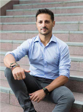
Dr Carmine Gentile.
“We’re developing an approach that uses biological material – in this case, cells taken from the same patient – to generate living heart tissue to replace the damaged heart tissue,” Dr Gentile says.
The human heart is made up of contracting muscle cells with blood vessels providing oxygen and nutrients to the areas within the muscle wall. Dr Gentile and his team realised that to give the newly grown heart cells the best chance of being accepted into the patient’s existing heart tissue, they’d have to mimic the heart’s own microenvironment. Despite a challenging process, they have been able to do just that.
They’ve called the combination of human heart cells grown in the laboratory ‘cardiac spheroids’, or the more graspable ‘mini hearts’.
The broad concept is not new. Other researchers around the world have managed to 3D print alternate layers of blood cells and muscle cells. What makes Dr Gentile’s approach unique is that the mini hearts more fully integrate all the cell types present in the human heart, including preformed blood vessels. These cells are used as bio-ink in a bioprinter that was custom-made for Dr Gentile and his team.
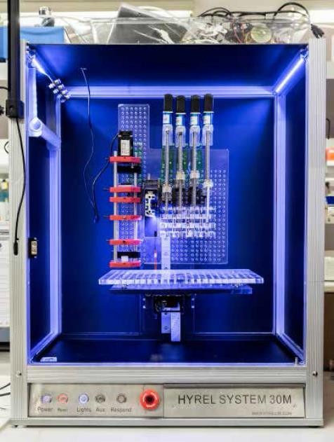
Professor Zreiqat's team uses this machine to print bone substitute.
The bioprinter, called ‘Reggie’ by Dr Gentile’s students, from the name of the Spanish company that built it, has a nozzle that sets down a layer of water-based hydrogel, and another nozzle that applies the cell-containing bio-ink. The resulting tissue mimics the mechanical and physical properties of the human heart and, since each patient has different requirements, a computer can vary the geometry of the multiple layers.
Dr Gentile’s mini heart research also offers the promise of an alternative to testing drugs on animals or with standard cell cultures. In a 2017 study, Dr Gentile and his team found that since mini hearts can be made with a patient’s own cells, they can be used to identify potential side effects that a person may experience from particular heart medications.
While Dr Gentile is mending hearts, Professor Hala Zreiqat is building bones. Born in Jordan, she’s a powerhouse of energy and ideas, and her achievements saw her win the 2018 NSW Premier’s Award for Woman of the Year. As head of the University’s Biomaterials and Tissue Engineering Research Unit, she has developed a technique of 3D printing ceramic bone so it acts as a scaffold that contains all the ingredients needed for the body to foster bone growth at the site of the defect.
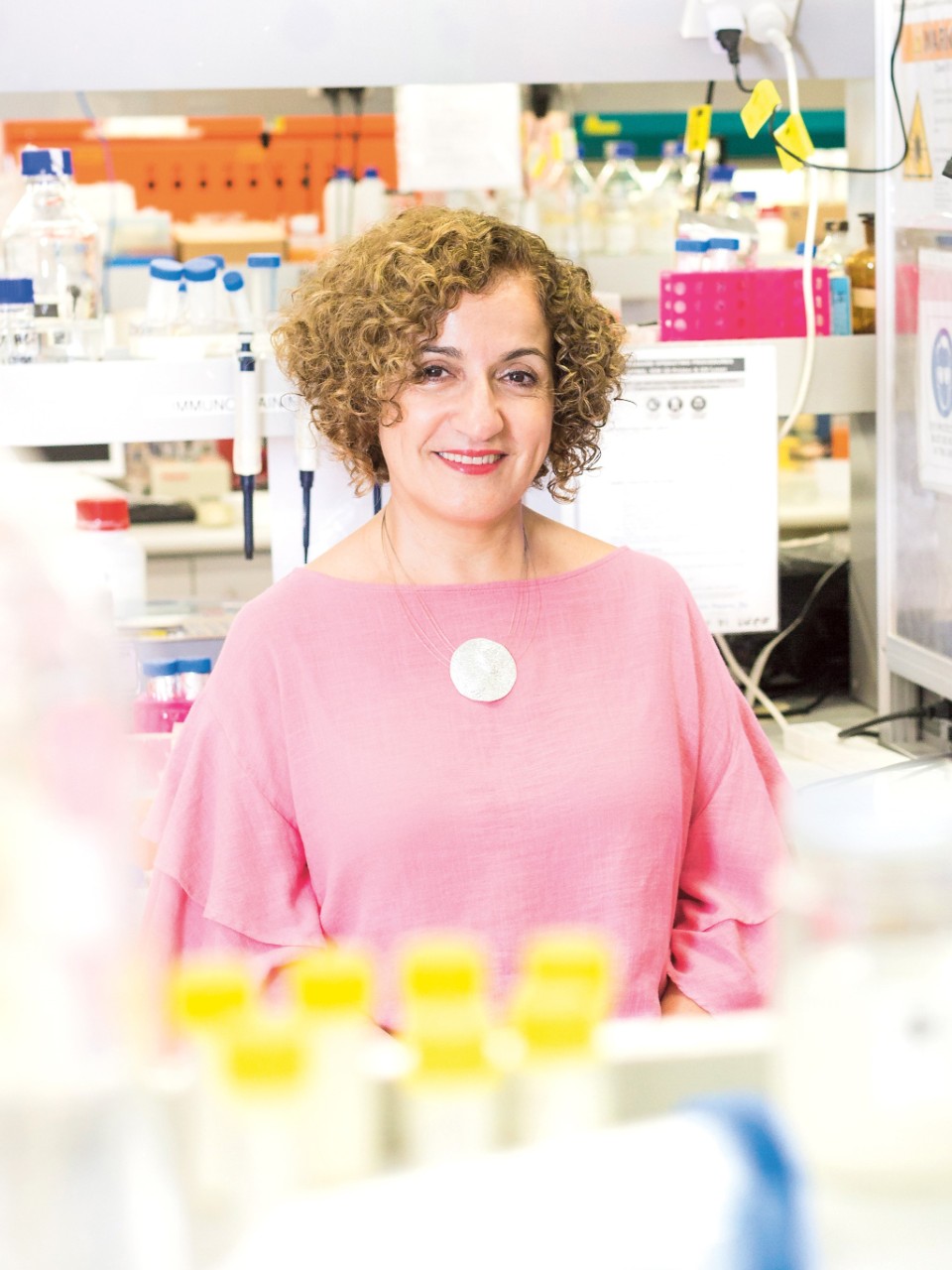
Professor Hala Zreiqat and her team draw knowledge from across the University community to contribute to leading-edge medicine.
Bone is the most transplanted substance in medicine, with loss or damage resulting from accidents, disease or developmental issues. Current treatments require either grafting from a secondary site, which is problematic when there has been large bone loss, or inserting metal implants which frequently need replacing as the body changes over time.
“What we’re working towards is taking a CT scan of the bone defect and feeding it straight into the printing machine, which hopefully would be sitting next to that operating theatre,” Professor Zreiqat says. “This is where the uniqueness of our material and discovery comes in. You can design any shape or size, so it can be applied to a really large or small bone defect.”
Healthy bone undergoes a constant process of renewal and bone growth. The bone substitute developed by Professor Zreiqat’s team incorporates ‘smart materials’ which contain trace elements and nanoparticles designed to promote bone growth. The non-toxic ceramic is porous, so blood and nutrients can penetrate it. Over time, the ceramic degrades as it is replaced by new bone.
You can design any shape or size, so it can be applied to a really large or small bone defect.
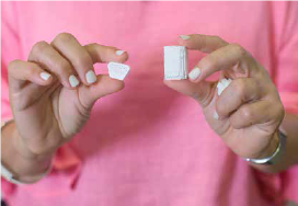
While real bone can be in short supply, Professor Zreiqat points out that 3D printed bone is made from materials that are plentiful.
A major challenge in producing ceramics suitable for bone replacement is matching the impressive load-bearing and shock absorption of actual bone. To approach this and other problems that have arisen during research, Professor Zreiqat encourages cross-collaboration between a wide range of disciplines.
Her team includes material scientists, cell and molecular biologists, chemical engineers, physicists and clinicians. In the future, she also expects to work with designers and architects. With four patents on board, and clinical trials expected to begin in two years, her interdisciplinary focus is paying off.
“Science and discoveries are all built around problems you don’t know the answer to,” she says. “So we draw expertise from each other to develop something new. For example, in our approach to materials, we thought about what would happen if we changed the architecture of that material? Working with the mathematical modelling people, we found that just by changing the architecture, you can significantly affect the quality and the type of bond that forms with the body. It’s big, right?
“I always think of the first coffee house that was opened in England back in the 1650s. When it opened, people started getting together from different disciplines to interact, and that’s where innovation and discovery started to happen in England. A multidisciplinary focus fosters an environment for innovation.”
To learn more about these ground-breaking ideas or help advance the work, call Sallie May on +61 2 8627 8818 or email the Sydney Development Fund.
Written by Rebekah Hayden
Photography by Louise Cooper
