Microscope that gets to the heart of matter
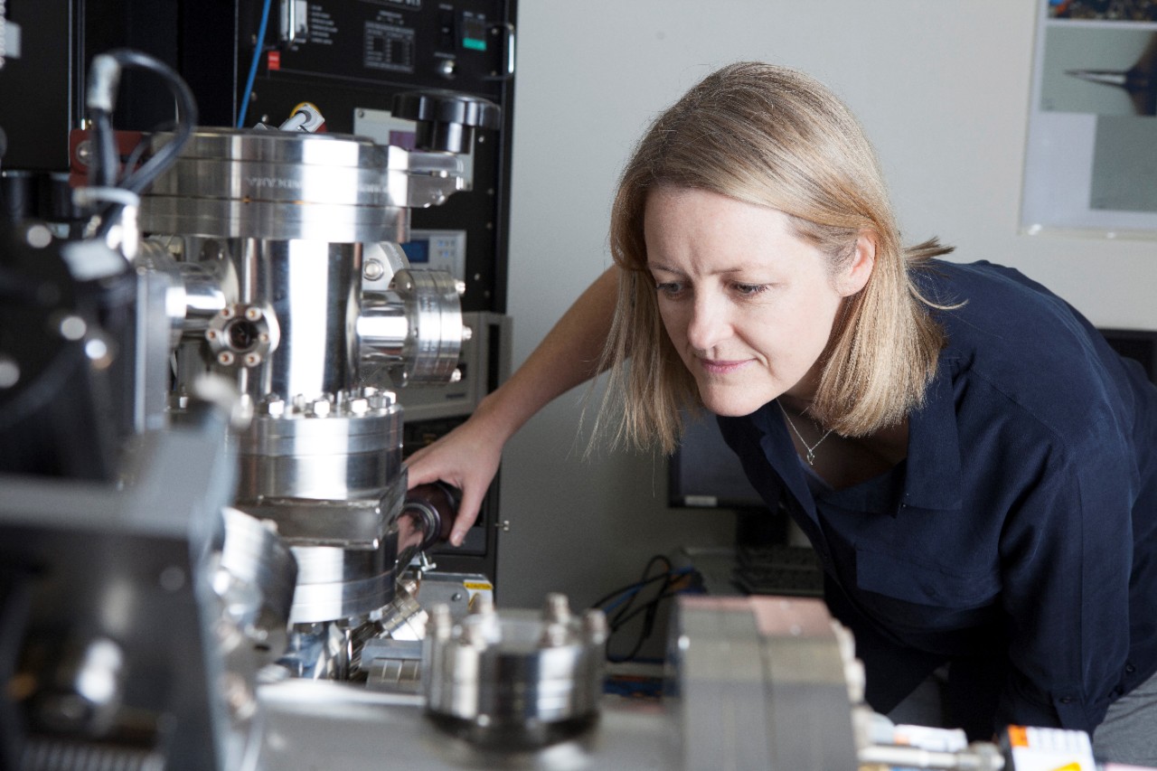
Professor Julie Cairney using an atom probe at the University of Sydney.
Two Nobel Laureates and the NSW Chief Scientist & Engineer have officially unveiled a vital piece of scientific infrastructure at the University of Sydney at a special event on Tuesday, 11 September 2018.
Professor Julie Cairney, director of Sydney Microscopy & Microanalysis, will use the multimillion-dollar microscope to improve the materials used to build life-saving stents that are inserted during heart surgery into patient’s aortas, saving many Australian lives each year.
“The work we will do with this machine has the potential to make a huge difference in the world. It will certainly have a transformational effect on my research,” Professor Cairney said.
The Thermo Fisher Themis-Z transmission electron microscope (TEM) has the highest resolution of any microscope in Australia. Its addition to the University of Sydney provides researchers with unparalleled access to the mysteries of the atomic structure of materials.
The 4.5-metre tall microscope is housed in the purpose-built $150 million Sydney Nanoscience Hub in a room that is shielded from electromagnetic interference and ‘floats’ architecturally independent from the building to minimise vibrations.
Speaking at the launch, the Vice-Chancellor and Principal, Dr Michael Spence, said: "This is just one machine in our community but it is really a contribution to scientific discourse across the world. It is through this kind of collaboration that we can meet our global challenges."
He said vision, hard work and collaboration will yield results through the science this instrument can facilitate.
The winner of the 2018 Eureka Prize for Leadership in Innovation and Science, Professor Thomas Maschmeyer, said the new machine will be a boon to his ground-breaking research into renewables.
“Defects in functional materials are often critical to performance, be that mechanical, electronic, optical or chemical. The new instrument will allow us to probe active sites, integral for catalysis or energy storage, with atomic clarity,” Professor Maschmeyer said.
Working with Cook Medical Australia, Professor Cairney, from the School of Aerospace, Mechanical and Mechatronic Engineering, is working on new materials that will allow doctors to better see and position the stents during the insertion procedure.
The new microscope will provide essential information about the structure and stability of these materials, allowing her team to be more effective in their work.
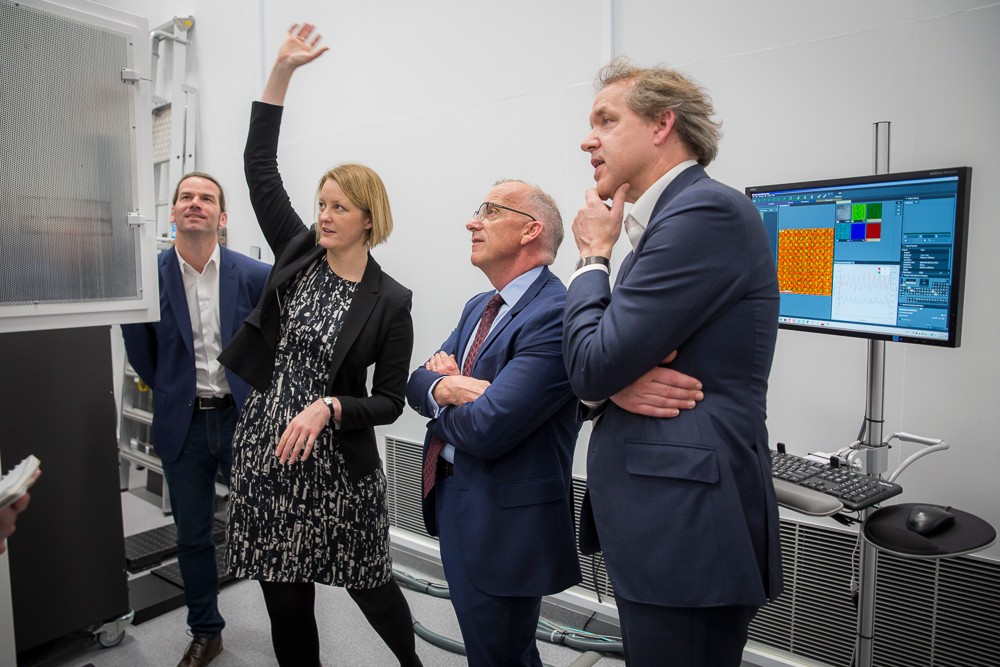
Professor Julie Cairney explains how the microscope works to Vice-Chancellor Dr Michael Spence (second from right) and Deputy Vice-Chancellor (Research) Professor Duncan Ivision (right). With them is Dr Magnus Garbrecht, who operates the TEM. Photo Jayne Ion
The TEM is the only such microscope in Australia that is “monochromated” and “double corrected”. This means it can simultaneously analyse the atomic structure and the spectral nature of materials.
The resolution of this technique is breathtaking. The machine can obtain images with resolution better than 0.06 billionths of a metre (0.06 nanometres). That is about 10 times smaller than the distance between silicon atoms or five times smaller than the distance between carbon atoms in diamond.
“With this device, we not only get to see the atomic structure but simultaneously analyse chemical information, such as inter-atomic forces, which is a huge advantage in materials science. The research application space is truly vast,” Professor Cairney said.
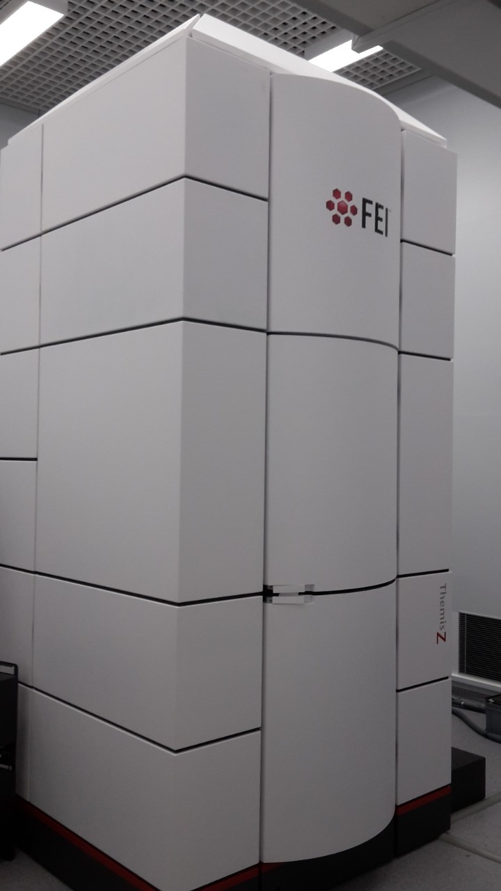
The aberration-corrected transmission electron microscope in the Sydney Nanoscience Hub.
The microscope, which is available for use by industry, has applications in geosciences, mining, chemical and advanced manufacturing industries. It is critical for the design of semiconductor structures such as those fabricated in cleanrooms at the Research and Prototype Foundry in the Sydney Nanoscience Hub.
Deputy Vice-Chancellor (Research), Professor Duncan Ivison, said: “This launch represents an important advance in Australia’s national capability in electron microscopy. This is a major milestone in the University’s research strategy, which places emphasis on world-class, University-wide infrastructure.”
The new NSW Chief Scientist & Engineer, Professor Hugh Durrant-Whyte launched the device in his first week of official functions alongside two Nobel Laureates in Chemistry who are visiting Sydney for the 19th International Microscopy Congress, which is jointly hosted by the University of Sydney and UNSW.
Columbia University’s Professor Joachim Frank was awarded a Nobel Prize last year for developing another innovative type of transmission electron microscopy, known as cryo-TEM, which is able to determine biological structures in solution to high resolution. The University of Sydney will soon acquire a cryo-TEM to add to our impressive array of scientific infrastructure.
"I want to emphasise the role of scientific infrastructure," Professor Frank said at the launch. "I've done most of my work computationally - and that data had to come from somewhere from people who can operate these microscopes.
"It is from efforts such as what I see happening here at Sydney where this happens. This really is a milestone for your institution."
Joining Professor Frank was Technion University Israel’s Professor Dan Shechtman. He was awarded the 2011 Nobel Prize for the discovery of quasicrystals, a highly unusual and complicated form of matter.
"You have fantastic equipment here," Professor Shechtman said. "But the questions are: where are the problems to solve? Will these wonderful pictures bring you new science? What do they mean for the real questions you want to solve.
"The most important thing is the man or woman behind the science. Developing the human capacity to use these instruments is the important job, almost as important as solving the scientific questions themselves."
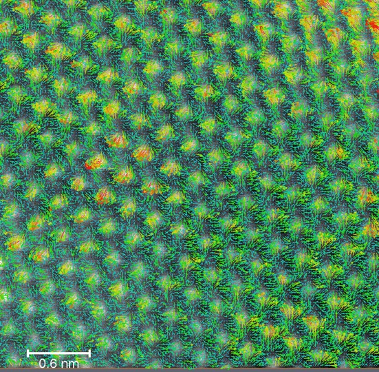
Differential phase contrast in scanning transmission electron microscopy mode reveals the electric field vectors around single magnesium atoms acting as dopants in scandium nitride. Image by Magnus Garbrecht
