
University team develops antibacterial surfaces for bone implants
A University of Sydney team in collaboration with the University Medical Centre Utrecht has found a solution to the infection-related rejection of implants, including knees and hips, that currently experience a 10 percent rejection rate and 2 percent infection rate, by developing a silver nanoparticle plasma coating.
In Australia alone, close to a million hip and knee replacement operations have been performed since 1999.
Each year, another 90,000 devices are surgically inserted at a cost of around $1 billion.
Around 10 percent of these operations require revision surgeries due to problems associated with poor bone integration and infection.
The rate of infection is around 1-2 percent, however the consequence are catastrophic as these infections are untreatable and require invasive removal of the implant followed by extensive antibiotic treatment, and re-implantation. The associated costs and morbidity are extremely high.
This new class of antimicrobial interfaces will benefit patients suffering from bone fracture, osteoporosis, and bone cancer.
The University of Sydney’s Dr Behnam Akhavan from the Applied Plasma and Surface Engineering Research Group in the Faculty of Engineering and Information Technologies in collaboration with the University Medical Centre Utrecht in the Netherlands, has developed a simple plasma-based technology for the fabrication of antimicrobial silver-functionalized coatings that are not toxic to mammalian cells. The research has created a new biomaterial surface that retains antibiotic silver nanoparticles over the long-term without being toxic to the surrounding tissue.
“Silver nanoparticles are naturally antibacterial, so having them on an implant surface, makes the implant antimicrobial. However, large doses of silver released from a surface to the surrounding tissue are harmful and toxic to the living tissue.
“A strategy is required to fix these silver nanoparticles on the surface, where they are needed to kill colonising bacteria, and at the same time prevent their release to the surrounding tissue,” said Dr Akhavan.
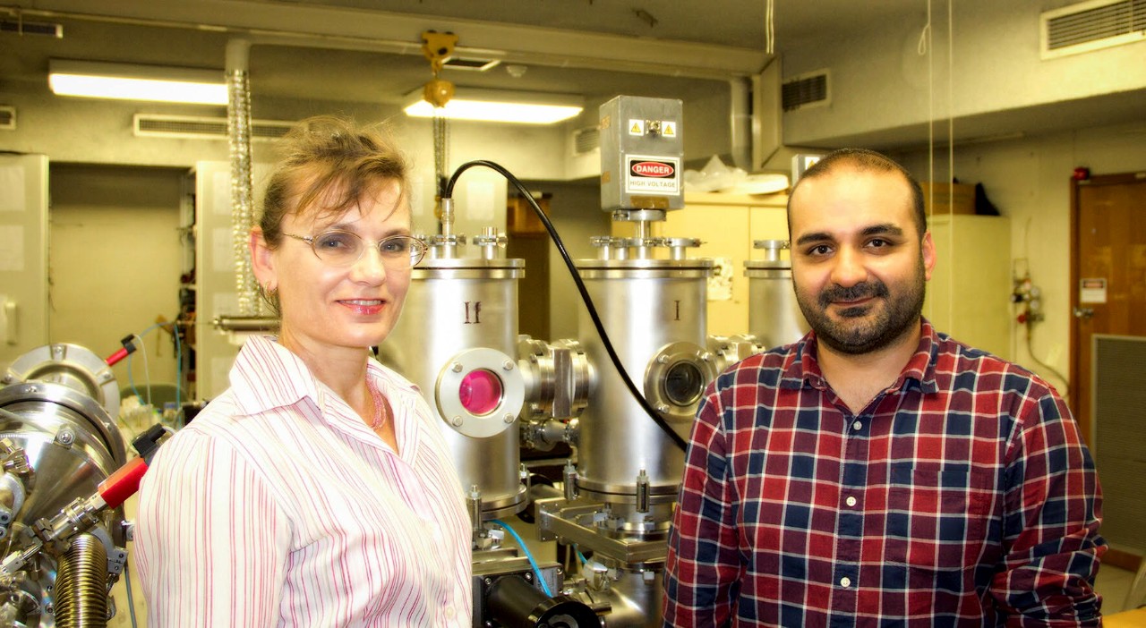
Professor Marcela Bilek and Dr Behnam Akhavan
The University of Sydney plasma-based technology provided the solution in the form of irreversible (permanent) attachment of silver nanoparticles on implant surfaces, so they show local antimicrobial activity only at the surface without affecting the surrounding tissue.
“Antibiotic resistance, that is one of the biggest threats to humans, is associated with conventional strategies that rely on the release of antibiotics from implant surface coatings, explains Dr Akhavan
A major impact of this research is the creation of orthopaedic implants which prevent infection without the risk of creating antibiotic resistant strains of bacteria as occurs with materials that release antibiotics.”
The developed coatings also allow the coupling of biomolecules to facilitate accelerated integration with natural bone tissue.
This industry-scalable approach allows for a local antimicrobial effect at the surface of the device, whilst minimising cytotoxicity associated with the release of silver into adjacent host tissue and presenting no risk of creating antibiotic resistant strains of bacteria.
This exciting development, coupled with our coating’s ability to irreversibly attach bone-signalling biomolecules, allows us to create biomedical implant surfaces that simultaneously repel infection and integrate with native body tissues.
The development opens up the possibility of fabricating a new generation of antimicrobial bone-implantable devices with improved tissue-implant integration.
"This exciting development, coupled with our coating’s ability to irreversibly attach bone-signalling biomolecules, allows us to create biomedical implant surfaces that simultaneously repel infection and integrate with native body tissues," says Professor Marcela Bilek, Sydney Nano, Charles Perkins Centre, and Faculty of Science.
“This new class of antimicrobial interfaces will benefit patients suffering from bone fracture, osteoporosis, and bone cancer.” says Dr Behnam Akhavan
“The next stage of the research is to coat patient-specific porous 3D structures created by 3D printing of titanium by our collaborators at UMC Utrecht, where subsequent implant in-vivo testing will be performed.
“We hope to conduct clinical trials by attracting industry support and funding. This is an incredibly exciting project with the potential for real impact. Our partnership with UMC Utrecht demonstrates the power of multi-disciplinary collaboration across borders to tackle seemingly intractable problems,” concluded Dr Akhavan.
The new study was published in the Journal of Materials Chemistry B.