Radical call to overhaul leaf biology models using 3D imaging
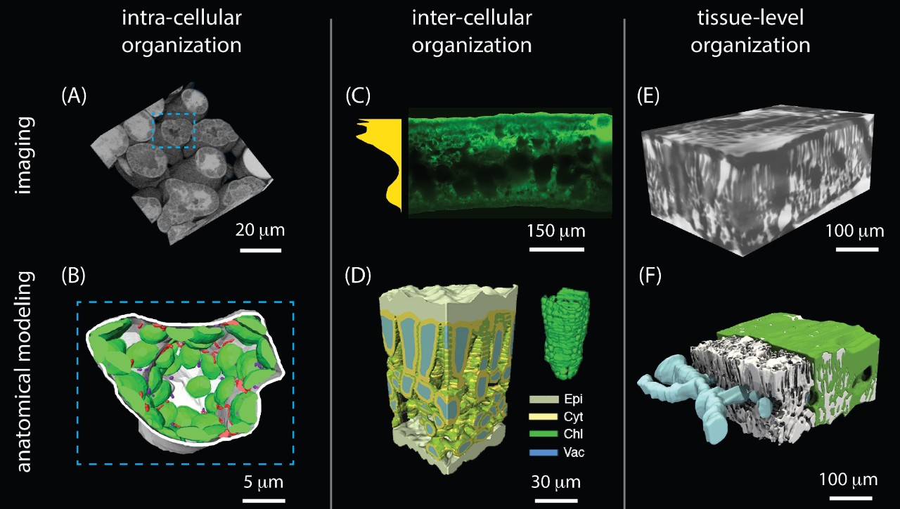
3D imaging can help us understand the complex processes inside leaves. From the paper we see images of sunflower, wheat and tomato leaves.
The field of plant science is in the process of being profoundly transformed by new imaging and modelling technologies. These tools are allowing scientists to peer inside the leaf with a clarity and resolution inconceivable a generation ago.
In an article published this month, a team of Australian and international scientists demonstrated how three-dimensional (3D) imaging can now reproduce the inner reality of the leaf, including the dynamic carbon and water exchange processes.

Professor Margaret Barbour.
Professor Margaret Barbour from the School of Life and Environmental Sciences at the University of Sydney said: “In this article, we show the huge potential that embracing 3D complexity can have in improving our understanding of leaves at multiple levels of biological organisation, including harnessing the knowledge to improve the photosynthetic performance of crops.
“It is a bit like being able to walk inside the leaf, instead of looking at it squashed in two dimensions,” said Professor Barbour, who is also an Associate investigator at the ARC Centre of Excellence for Translational Photosynthesis (CoETP).
A co-author of the paper, published in Trends in Plant Science, is Professor Graham Farquhar from the Australian National University. Professor Farquhar received the Kyoto Prize in 2017 and the Prime Minister's Prize for Science in 2015 in recognition for his work in developing our understanding of photosynthesis. He was also Professor Barbour's PhD supervisor.
Professor John Evans, from the Research School of Biology at The Australian National University, and one of the authors of the research, said that although leaves and plant cells are three dimensional, plant biologists use highly simplified 1D or 2D models, evading the difficult, confounding, and beautiful 3D reality.
3D image of wheat mesophyll.
“The leaf is an amazingly complex landscape, where water and gases flow in many directions depending on variables such as temperature, light quality and wind. 3D images give you an understanding of what is really happening,” said Professor Evans, who is a CoETP Chief investigator
"These technologies make it possible to answer very interesting questions, some of which have eluded scientists for many years,” he said.
The images are created from biological specimens, by integrating 2D leaf measurements to create 3D volumes and surfaces. The 3D representation enables an anatomically correct basis for modelling and biophysical simulations to provide a dynamic view of the processes inside plant cells and tissues.
The scientists predict that using a collaborative approach, they will be able to answer, within the next decade, outstanding questions about how the 3D special arrangement of organelles, cells and tissues affects photosynthesis and transpiration.
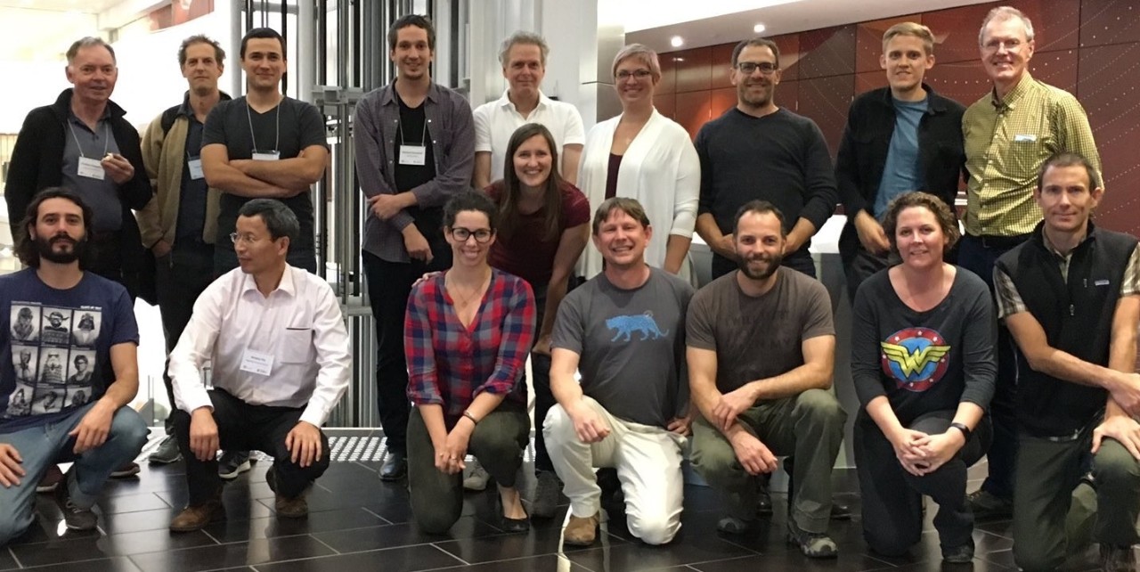
Meeting at the University of Sydney of the 3D leaf imaging group.
The work is an international collaboration involving researchers from Yale, Harvard, UC Davis, UCLA, Wageningen, University of Idaho, Western Sydney University and University of Queensland, supported by the University of Sydney and the ARC Centre for Translational Photosynthesis, whose lead node is located at ANU.
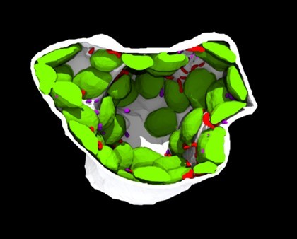
Inside a wheat cell
3D rendering inside a wheat mesophyll cell showing chloroplasts
