
Missing piece identified for lower-cost, high-quality MRI scans: study
Researchers from the University of Sydney in Australia and Massachusetts General Hospital in the United States have developed a new technique involving nanoparticles to improve the image quality of medical scans of portable low-cost Magnetic Resonance Imaging (MRI) machines. The development can help improve access to diagnostic imaging worldwide.
The study was published in the journal Science Advances.
Recently a new generation of portable, low-cost MRI scanners have had an increasing presence in hospitals. These machines are more versatile in medical settings compared to a standard MRI machine.
For instance, in the last six months, portable MRI scanners have been used to diagnose strokes and to monitor the neurological health of comatose COVID-19 patients, all while at the bedside.
However, the trade-off for this type of machine has been the quality and resolution of the medical scans. While these portable scanners operating at low magnetic field can be used to diagnose many conditions, the images are not as detailed as a standard MRI scan.
Using nanoparticles to improve image quality in portable MRIs
Nanoparticles key to technique
To improve the image quality of the portable MRI machines, University of Sydney researchers investigated a new technique of using nanoparticles to improve the image contrast in MRI images. The team of researchers included Dr David Waddington, Professor Zdenka Kuncic from the University of Sydney Nano Institute and Charles Perkins Centre, with PhD students Thomas Boele and Richard Maschmeyer, .
“We tested superparamagnetic iron oxide nanoparticles (SPIONs) as a contrast agent – they essentially amplify magnetic fields,” said Dr Waddington, lead author and Cancer Institute NSW Early Career Fellow from the University’s ACRF Image X Institute and Faculty of Medicine and Health.
“At low magnetic fields, these iron oxide nanoparticles are 3000 times more magnetic than conventional MRI contrast agents, which means they work really well in the lower magnetic fields of portable MRI scanners.”
The before and after images of the study in mice show a striking difference with organs clearly identified following the administration of SPIONs.
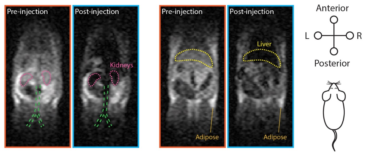
Before and after MRI images in mice with organs clearly identified following the administration of SPIONs in the study.
Making MRI scanners more affordable has been a long-term goal of the study’s co-author Dr Matthew Rosen, Director of the Low-field MRI and Hyperpolarized Media Laboratory at Massachusetts General Hospital.
Dr Rosen explained that low-field MRIs are growing in popularity, however an injectable contrast agent imaging technique that works similarly to those used in conventional MRI scanners is needed to improve the quality of images they produce. Doctors occasionally inject patients with a contrast agent based on the heavy metal gadolinium before performing a conventional MRI to enhance image quality, though concerns about long-term toxicity now limit that practice.
For gadolinium to be used with low-field MRI, a doctor would have to administer 1000 times more than the approved amount.
“SPIONs are safe and approved in the US and Europe for treating anaemia. While they need to be approved for specific use as contrast agents , doctors can use them now “off label” (they can be prescibed) with low-field MRI.
“We believe the combination of portable low-field MRIs and SPIONs will make low-cost MRI a reality.”
Dr Waddington and Professor Kuncic are also investigating the use of specially coated SPIONs that could allow MRI to be used for detecting malignant tumours.
Improving medical access
“Buying and installing an MRI scanner typically costs more than $2 million (AUD). This is why the machines are primarily found in imaging clinics and are unaffordable for medical centres in remote areas with small patient populations,” said Dr Waddington.
“Lowering the cost of MRI scanners could greatly expand the role of this powerful imaging technology in medicine by increasing the availability of scanning in emergency rooms, intensive-care units, and doctors’ offices.”
About the study:
Healthy mice were scanned with a home-made ultra-low field (ULF) MRI scanner (0.0065 T) in Dr Rosen’s lab, then injected with SPIONs and rescanned. A comparison of before and after injection images shows a striking difference, with kidneys, livers, and other organs glowing brightly following administration of SPIONs.
About MRI scanners:
The cost of an MRI scanner is largely driven by its superconducting magnet, the stronger the magnetic field it produces, the more expensive the machine. A typical MRI machine generates a magnetic field of 1.5 Tesla (T), though increasingly machines reach 3 T. However, a new generation of portable “low-field” MRI scanners that operate at 0.064 T and cost around $100,000 (AUD) has recently become available.
Declaration:
This work was supported by the University of Sydney Nano Institute. Matthew Rosen acknowledges funding from the Advanced Research Projects Agency-Energy (ARPA-E), U.S. Department of Energy. David Waddington is supported by a Cancer Institute of NSW Early Career Fellowship.
Zdenka Kuncic acknowledges funding from an Australian Academy of Technological Sciences and Engineering (ATSE) Global Connections Fund Bridging Grant.
Matthew Rosen is a cofounder of Hyperfine Research Inc., a manufacturer of portable MRI scanners. Zdenka Kuncic, Matthew Rosen and David Waddington are inventors on a patent application relating to the use of SPIONs as low-field contrast agents.